
BD HorizonTM BUV737 Rat Anti-Mouse CD25 (IL-2 Receptor alpha)
569602 · 25 ug
BDB Bioscience · Cat: 569602
Reach 3+ verified distributors
Manufacturer product page
bdbiosciences.com/en-us/products/reagents/flow-cytometry-rea...Description
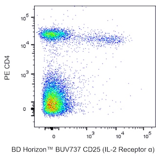
Flow cytometric analysis of CD25 IL-2 Receptor alpha expression on unstimulated and stimulated Mouse splenic leukocytes. Left and Middle Plots: Mouse splenic leukocytes were preincubated with Purified Rat Anti-Mouse CD16/CD32 antibody Mouse BD Fc Block Cat. No. 553142 . The cells were then stained with PE Rat Anti-Mouse CD4 antibody Cat. No. 553049 and with either BD Horizon BUV737 Rat IgG1, kappa Isotype Control Cat. No. 612770; Left Plot or BD Horizon BUV737 Rat Anti-Mouse CD25 IL-2 Receptor alpha antibody Cat. No. 569600/569601; Middle Plot at 0.5 mug/test. DAPI 4,6-Diamidino-2-Phenylindole, Dihydrochloride Solution Cat. No. 564907 was added to cells right before analysis. The bivariate pseudocolor density plot showing the correlated expression of CD25 IL-2 Receptor alpha or Ig Isotype control staining versus CD4 was derived from DAPI negative-gated events with the forward and side light-scatter characteristics of viable leukocytes. Right Plot: Mouse splenic leukocytes were stimulated for 3 days with Concanavalin A ConA . The cells were preincubated with Purified Rat Anti-Mouse CD16/CD32 antibody and then stained with either BD Horizon BUV737 Rat IgG1, kappa Isotype Control dashed line histogram or BD Horizon BUV737 Rat Anti-Mouse CD25 IL-2 Receptor alpha antibody solid line histogram at 0.5 mug/test. The fluorescence histogram showing CD25 IL-2 Receptor alpha expression or Ig Isotype control staining was derived from DAPI negative-events with the forward and side light-scatter characteristics of viable lymphoblasts. Flow cytometry and data analysis were performed using a BD LSRFortessa X-20 Flow Cytometer System and FlowJo software. Data shown on this Technical Data Sheet are not lot specific. Flow cytometric analysis of CD25 IL-2 Receptor alpha expression on unstimulated and stimulated Mouse splenic leukocytes. Left and Middle Plots: Mouse splenic leukocytes were preincubated with Purified Rat Anti-Mouse CD16/CD32 antibody Mouse BD Fc Block Cat. No. 553142 . The cells were then stained with PE Rat Anti-Mouse CD4 antibody Cat. No. 553049 and with either BD Horizon BUV737 Rat IgG1, kappa Isotype Control Cat. No. 612770; Left Plot or BD Horizon BUV737 Rat Anti-Mouse CD25 IL-2 Receptor alpha antibody Cat. No. 569600/569601; Middle Plot at 0.5 mug/test. DAPI 4,6-Diamidino-2-Phenylindole, Dihydrochloride Solution Cat. No. 564907 was added to cells right before analysis. The bivariate pseudocolor density plot showing the correlated expression of CD25 IL-2 Receptor alpha or Ig Isotype control staining versus CD4 was derived from DAPI negative-gated events with the forward and side light-scatter characteristics of viable leukocytes. Right Plot: Mouse splenic leukocytes were stimulated for 3 days with Concanavalin A ConA . The cells were preincubated with Purified Rat Anti-Mouse CD16/CD32 antibody and then stained with either BD Horizon BUV737 Rat IgG1, kappa Isotype Control dashed line histogram or BD Horizon BUV737 Rat Anti-Mouse CD25 IL-2 Receptor alpha antibody solid line histogram at 0.5 mug/test. The fluorescence histogram showing CD25 IL-2 Receptor alpha expression or Ig Isotype control staining was derived from DAPI negative-events with the forward and side light-scatter characteristics of viable lymphoblasts. Flow cytometry and data analysis were performed using a BD LSRFortessa X-20 Flow Cytometer System and FlowJo software. Data shown on this Technical Data Sheet are not lot specific. Flow cytometric analysis of CD25 IL-2 Receptor alpha expression on unstimulated and stimulated Mouse splenic leukocytes. Left and Middle Plots: Mouse splenic leukocytes were preincubated with Purified Rat Anti-Mouse CD16/CD32 antibody Mouse BD Fc Block Cat. No. 553142 . The cells were then stained with PE Rat Anti-Mouse CD4 antibody Cat. No. 553049 and with either BD Horizon BUV737 Rat IgG1, kappa Isotype Control Cat. No. 612770; Left Plot or BD Horizon BUV737 Rat Anti-Mouse CD25 IL-2 Receptor alpha antibody Cat. No. 569600/569601; Middle Plot at 0.5 mug/test. DAPI 4,6-Diamidino-2-Phenylindole, Dihydrochloride Solution Cat. No. 564907 was added to cells right before analysis. The bivariate pseudocolor density plot showing the correlated expression of CD25 IL-2 Receptor alpha or Ig Isotype control staining versus CD4 was derived from DAPI negative-gated events with the forward and side light-scatter characteristics of viable leukocytes. Right Plot: Mouse splenic leukocytes were stimulated for 3 days with Concanavalin A ConA . The cells were preincubated with Purified Rat Anti-Mouse CD16/CD32 antibody and then stained with either BD Horizon BUV737 Rat IgG1, kappa Isotype Control dashed line histogram or BD Horizon BUV737 Rat Anti-Mouse CD25 IL-2 Receptor alpha antibody solid line histogram at 0.5 mug/test. The fluorescence histogram showing CD25 IL-2 Receptor alpha expression or Ig Isotype control staining was derived from DAPI negative-events with the forward and side light-scatter characteristics of viable lymphoblasts. Flow cytometry and data analysis were performed using a BD LSRFortessa X-20 Flow Cytometer System and FlowJo software. Data shown on this Technical Data Sheet are not lot specific. Show More Flow cytometric analysis of CD25 IL-2 Receptor alpha expression on unstimulated and stimulated Mouse splenic leukocytes. Left and Middle Plots: Mouse splenic leukocytes were preincubated with Purified Rat Anti-Mouse CD16/CD32 antibody Mouse BD Fc Block Cat. No. 553142 . The cells were then stained with PE Rat Anti-Mouse CD4 antibody Cat. No. 553049 and with either BD Horizon BUV737 Rat IgG1, kappa Isotype Control Cat. No. 612770; Left Plot or BD Horizon BUV737 Rat Anti-Mouse CD25 IL-2 Receptor alpha antibody Cat. No. 569600/569601; Middle Plot at 0.5 mug/test. DAPI 4,6-Diamidino-2-Phenylindole, Dihydrochloride Solution Cat. No. 564907 was added to cells right before analysis. The bivariate pseudocolor density plot showing the correlated expression of CD25 IL-2 Receptor alpha or Ig Isotype control staining versus CD4 was derived from DAPI negative-gated events with the forward and side light-scatter characteristics of viable leukocytes. Right Plot: Mouse splenic leukocytes were stimulated for 3 days with Concanavalin A ConA . The cells were preincubated with Purified Rat Anti-Mouse CD16/CD32 antibody and then stained with either BD Horizon BUV737 Rat IgG1, kappa Isotype Control dashed line histogram or BD Horizon BUV737 Rat Anti-Mouse CD25 IL-2 Receptor alpha antibody solid line histogram at 0.5 mug/test. The fluorescence histogram showing CD25 IL-2 Receptor alpha expression or Ig Isotype control staining was derived from DAPI negative-events with the forward and side light-scatter characteristics of viable lymphoblasts. Flow cytometry and data analysis were performed using a BD LSRFortessa X-20 Flow Cytometer System and FlowJo software. Data shown on this Technical Data Sheet are not lot specific. Show More Flow cytometric analysis of CD25 IL-2 Receptor alpha expression on unstimulated and stimulated Mouse splenic leukocytes. Left and Middle Plots: Mouse splenic leukocytes were preincubated with Purified Rat Anti-Mouse CD16/CD32 antibody Mouse BD Fc Block Cat. No. 553142 . The cells were then stained with PE Rat Anti-Mouse CD4 antibody Cat. No. 553049 and with either BD Horizon BUV737 Rat IgG1, kappa Isotype Control Cat. No. 612770; Left Plot or BD Horizon BUV737 Rat Anti-Mouse CD25 IL-2 Receptor alpha antibody Cat. No. 569600/569601; Middle Plot at 0.5 mug/test. DAPI 4,6-Diamidino-2-Phenylindole, Dihydrochloride Solution Cat. No. 564907 was added to cells right before analysis. The bivariate pseudocolor density plot showing the correlated expression of CD25 IL-2 Receptor alpha or Ig Isotype control staining versus CD4 was derived from DAPI negative-gated events with the forward and side light-scatter characteristics of viable leukocytes. Right Plot: Mouse splenic leukocytes were stimulated for 3 days with Concanavalin A ConA . The cells were preincubated with Purified Rat Anti-Mouse CD16/CD32 antibody and then stained with either BD Horizon BUV737 Rat IgG1, kappa Isotype Control dashed line histogram or BD Horizon BUV737 Rat Anti-Mouse CD25 IL-2 Receptor alpha antibody solid line histogram at 0.5 mug/test. The fluorescence histogram showing CD25 IL-2 Receptor alpha expression or Ig Isotype control staining was derived from DAPI negative-events with the forward and side light-scatter characteristics of viable lymphoblasts. Flow cytometry and data analysis were performed using a BD LSRFortessa X-20 Flow Cytometer System and FlowJo software. Data shown on this Technical Data Sheet are not lot specific. Flow cytometric analysis of CD25 IL-2 Receptor alpha expression on unstimulated and stimulated Mouse splenic leukocytes. Left and Middle Plots: Mouse splenic leukocytes were preincubated with Purified Rat Anti-Mouse CD16/CD32 antibody Mouse BD Fc Block Cat. No. 553142 . The cells were then stained with PE Rat Anti-Mouse CD4 antibody Cat. No. 553049 and with either BD Horizon BUV737 Rat IgG1, kappa Isotype Control Cat. No. 612770; Left Plot or BD Horizon BUV737 Rat Anti-Mouse CD25 IL-2 Receptor alpha antibody Cat. No. 569600/569601; Middle Plot at 0.5 mug/test. DAPI 4,6-Diamidino-2-Phenylindole, Dihydrochloride Solution Cat. No. 564907 was added to cells right before analysis. The bivariate pseudocolor density plot showing the correlated expression of CD25 IL-2 Receptor alpha or Ig Isotype control staining versus CD4 was derived from DAPI negative-gated events with the forward and side light-scatter characteristics of viable leukocytes. Right Plot: Mouse splenic leukocytes were stimulated for 3 days with Concanavalin A ConA . The cells were preincubated with Purified Rat Anti-Mouse CD16/CD32 antibody and then stained with either BD Horizon BUV737 Rat IgG1, kappa Isotype Control dashed line histogram or BD Horizon BUV737 Rat Anti-Mouse CD25 IL-2 Receptor alpha antibody solid line histogram at 0.5 mug/test. The fluorescence histogram showing CD25 IL-2 Receptor alpha expression or Ig Isotype control staining was derived from DAPI negative-events with the forward and side light-scatter characteristics of viable lymphoblasts. Flow cytometry and data analysis were performed using a BD LSRFortessa X-20 Flow Cytometer System and FlowJo software. Data shown on this Technical Data Sheet are not lot specific.