
BD HorizonTM BV421 Goat Anti-Mouse Ig
563846 ·
BDB Bioscience · Cat: 563846
Reach 3+ verified distributors
Manufacturer product page
bdbiosciences.com/en-us/products/reagents/flow-cytometry-rea...Description
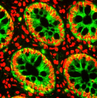
Flow cytometric analysis of CD3 expression on human peripheral blood lymphocytes and CD4 expression on rat splenic leucocytes. Far Left: Human whole blood was incubated with either no primary antibody dashed line histogram or Purified Mouse Anti-Human CD3 antibody Cat. No. 555330; solid line histogram . Human erythrocytes were lysed with BD PharmLyse Cat. No. 555899 . Middle Left: Rat splenic leucocytes were similarly incubated with either no antibody dashed line histogram or Purified Mouse Anti-Rat CD4 antibody Cat. No. 554835; solid line histogram . Both human and rat cells were washed and stained with BD Horizon BV421 Goat Anti-Mouse Ig antibody Cat. No. 563846 . The fluorescence histograms were derived from gated events with the forward and side light-scatter characteristics of viable human lymphocytes or rat leucocytes. Flow cytometric analysis was performed using a BD LSR II Flow Cytometer System. Immunofluorescent and immunohistochemical analysis of E-cadherin expression on human breast adenocarcinoma and large intestine. Middle Right: MCF-7 ATCC HTB-22 were fixed with BD Cytofix buffer Cat. No. 554655 , permeabilized with BD Phosflow Perm Buffer III Cat. No. 558050 , blocked, and stained with Purified Mouse anti-E-cadherin Cat. No. 610182 at 5 mug/mL. Second step reagent was BD Horizon BV421 Goat Anti-Mouse Ig at 1.25 mug/mL pseudo-colored green , captured on a Zeiss LSM 710 confocal microscope, merged using Image J Software. Far Right: Frozen sections of intestine were fixed with BD Cytofix buffer, permeabilized with 0.2 % Triton X-100, blocked, and stained with Purified Mouse anti-E-cadherin at 5 mug/mL. Second step reagent was BD Horizon BV421 Goat Anti-Mouse Ig at 1.25 mug/mL pseudo-colored green , captured on a BD Pathway 435 Cell Analyzer, merged using BD Attovision Software. For both samples, nuclei were counter-stained with DRAQ5 pseudo-colored red and slides were mounted with ProLong Gold. Flow cytometric analysis of CD3 expression on human peripheral blood lymphocytes and CD4 expression on rat splenic leucocytes. Far Left: Human whole blood was incubated with either no primary antibody dashed line histogram or Purified Mouse Anti-Human CD3 antibody Cat. No. 555330; solid line histogram . Human erythrocytes were lysed with BD PharmLyse Cat. No. 555899 . Middle Left: Rat splenic leucocytes were similarly incubated with either no antibody dashed line histogram or Purified Mouse Anti-Rat CD4 antibody Cat. No. 554835; solid line histogram . Both human and rat cells were washed and stained with BD Horizon BV421 Goat Anti-Mouse Ig antibody Cat. No. 563846 . The fluorescence histograms were derived from gated events with the forward and side light-scatter characteristics of viable human lymphocytes or rat leucocytes. Flow cytometric analysis was performed using a BD LSR II Flow Cytometer System. Immunofluorescent and immunohistochemical analysis of E-cadherin expression on human breast adenocarcinoma and large intestine. Middle Right: MCF-7 ATCC HTB-22 were fixed with BD Cytofix buffer Cat. No. 554655 , permeabilized with BD Phosflow Perm Buffer III Cat. No. 558050 , blocked, and stained with Purified Mouse anti-E-cadherin Cat. No. 610182 at 5 mug/mL. Second step reagent was BD Horizon BV421 Goat Anti-Mouse Ig at 1.25 mug/mL pseudo-colored green , captured on a Zeiss LSM 710 confocal microscope, merged using Image J Software. Far Right: Frozen sections of intestine were fixed with BD Cytofix buffer, permeabilized with 0.2 % Triton X-100, blocked, and stained with Purified Mouse anti-E-cadherin at 5 mug/mL. Second step reagent was BD Horizon BV421 Goat Anti-Mouse Ig at 1.25 mug/mL pseudo-colored green , captured on a BD Pathway 435 Cell Analyzer, merged using BD Attovision Software. For both samples, nuclei were counter-stained with DRAQ5 pseudo-colored red and slides were mounted with ProLong Gold. Show More Flow cytometric analysis of CD3 expression on human peripheral blood lymphocytes and CD4 expression on rat splenic leucocytes. Far Left: Human whole blood was incubated with either no primary antibody dashed line histogram or Purified Mouse Anti-Human CD3 antibody Cat. No. 555330; solid line histogram . Human erythrocytes were lysed with BD PharmLyse Cat. No. 555899 . Middle Left: Rat splenic leucocytes were similarly incubated with either no antibody dashed line histogram or Purified Mouse Anti-Rat CD4 antibody Cat. No. 554835; solid line histogram . Both human and rat cells were washed and stained with BD Horizon BV421 Goat Anti-Mouse Ig antibody Cat. No. 563846 . The fluorescence histograms were derived from gated events with the forward and side light-scatter characteristics of viable human lymphocytes or rat leucocytes. Flow cytometric analysis was performed using a BD LSR II Flow Cytometer System. Immunofluorescent and immunohistochemical analysis of E-cadherin expression on human breast adenocarcinoma and large intestine. Middle Right: MCF-7 ATCC HTB-22 were fixed with BD Cytofix buffer Cat. No. 554655 , permeabilized with BD Phosflow Perm Buffer III Cat. No. 558050 , blocked, and stained with Purified Mouse anti-E-cadherin Cat. No. 610182 at 5 mug/mL. Second step reagent was BD Horizon BV421 Goat Anti-Mouse Ig at 1.25 mug/mL pseudo-colored green , captured on a Zeiss LSM 710 confocal microscope, merged using Image J Software. Far Right: Frozen sections of intestine were fixed with BD Cytofix buffer, permeabilized with 0.2 % Triton X-100, blocked, and stained with Purified Mouse anti-E-cadherin at 5 mug/mL. Second step reagent was BD Horizon BV421 Goat Anti-Mouse Ig at 1.25 mug/mL pseudo-colored green , captured on a BD Pathway 435 Cell Analyzer, merged using BD Attovision Software. For both samples, nuclei were counter-stained with DRAQ5 pseudo-colored red and slides were mounted with ProLong Gold.