
BD HorizonTM BV480 Mouse Anti-Human CD279 (PD-1)
566112 · 100 Tests
BDB Bioscience · Cat: 566112
Reach 3+ verified distributors
Manufacturer product page
bdbiosciences.com/en-us/products/reagents/flow-cytometry-rea...Description
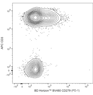
Two-color flow cytometric analysis of CD279 PD-1 expression on human peripheral blood lymphocytes. Human whole blood was stained with APC Mouse Anti-Human CD3 antibody Cat. No. 561810/555335 and either BD Horizon BV480 Mouse IgG1, kappa Isotype Control Cat. No. 565652; Left Plot or BD Horizon BV480 Mouse Anti-Human CD279 PD-1 antibody Cat. No. 566112/566175; Right Plot . Erythrocytes were lysed with BD FACS Lysing Solution Cat. No. 349202 . The two-color flow cytometric contour plot showing the correlated expression of CD279 PD-1 or Ig Isotype control staining versus CD3 was derived from gated events with the forward and side light-scatter characteristics of intact lymphocytes. Flow cytometric analysis was performed using a BD LSRFortessa Cell Analyzer System. Two-color flow cytometric analysis of CD279 PD-1 expression on human peripheral blood lymphocytes. Human whole blood was stained with APC Mouse Anti-Human CD3 antibody Cat. No. 561810/555335 and either BD Horizon BV480 Mouse IgG1, kappa Isotype Control Cat. No. 565652; Left Plot or BD Horizon BV480 Mouse Anti-Human CD279 PD-1 antibody Cat. No. 566112/566175; Right Plot . Erythrocytes were lysed with BD FACS Lysing Solution Cat. No. 349202 . The two-color flow cytometric contour plot showing the correlated expression of CD279 PD-1 or Ig Isotype control staining versus CD3 was derived from gated events with the forward and side light-scatter characteristics of intact lymphocytes. Flow cytometric analysis was performed using a BD LSRFortessa Cell Analyzer System. Two-color flow cytometric analysis of CD279 PD-1 expression on human peripheral blood lymphocytes. Human whole blood was stained with APC Mouse Anti-Human CD3 antibody Cat. No. 561810/555335 and either BD Horizon BV480 Mouse IgG1, kappa Isotype Control Cat. No. 565652; Left Plot or BD Horizon BV480 Mouse Anti-Human CD279 PD-1 antibody Cat. No. 566112/566175; Right Plot . Erythrocytes were lysed with BD FACS Lysing Solution Cat. No. 349202 . The two-color flow cytometric contour plot showing the correlated expression of CD279 PD-1 or Ig Isotype control staining versus CD3 was derived from gated events with the forward and side light-scatter characteristics of intact lymphocytes. Flow cytometric analysis was performed using a BD LSRFortessa Cell Analyzer System. Show More Two-color flow cytometric analysis of CD279 PD-1 expression on human peripheral blood lymphocytes. Human whole blood was stained with APC Mouse Anti-Human CD3 antibody Cat. No. 561810/555335 and either BD Horizon BV480 Mouse IgG1, kappa Isotype Control Cat. No. 565652; Left Plot or BD Horizon BV480 Mouse Anti-Human CD279 PD-1 antibody Cat. No. 566112/566175; Right Plot . Erythrocytes were lysed with BD FACS Lysing Solution Cat. No. 349202 . The two-color flow cytometric contour plot showing the correlated expression of CD279 PD-1 or Ig Isotype control staining versus CD3 was derived from gated events with the forward and side light-scatter characteristics of intact lymphocytes. Flow cytometric analysis was performed using a BD LSRFortessa Cell Analyzer System. Show More Two-color flow cytometric analysis of CD279 PD-1 expression on human peripheral blood lymphocytes. Human whole blood was stained with APC Mouse Anti-Human CD3 antibody Cat. No. 561810/555335 and either BD Horizon BV480 Mouse IgG1, kappa Isotype Control Cat. No. 565652; Left Plot or BD Horizon BV480 Mouse Anti-Human CD279 PD-1 antibody Cat. No. 566112/566175; Right Plot . Erythrocytes were lysed with BD FACS Lysing Solution Cat. No. 349202 . The two-color flow cytometric contour plot showing the correlated expression of CD279 PD-1 or Ig Isotype control staining versus CD3 was derived from gated events with the forward and side light-scatter characteristics of intact lymphocytes. Flow cytometric analysis was performed using a BD LSRFortessa Cell Analyzer System. Two-color flow cytometric analysis of CD279 PD-1 expression on human peripheral blood lymphocytes. Human whole blood was stained with APC Mouse Anti-Human CD3 antibody Cat. No. 561810/555335 and either BD Horizon BV480 Mouse IgG1, kappa Isotype Control Cat. No. 565652; Left Plot or BD Horizon BV480 Mouse Anti-Human CD279 PD-1 antibody Cat. No. 566112/566175; Right Plot . Erythrocytes were lysed with BD FACS Lysing Solution Cat. No. 349202 . The two-color flow cytometric contour plot showing the correlated expression of CD279 PD-1 or Ig Isotype control staining versus CD3 was derived from gated events with the forward and side light-scatter characteristics of intact lymphocytes. Flow cytometric analysis was performed using a BD LSRFortessa Cell Analyzer System.