
BD HorizonTM BV750 Mouse Anti-Human TNF
566359 ·
BDB Bioscience · Cat: 566359
Reach 3+ verified distributors
Manufacturer product page
bdbiosciences.com/en-us/products/reagents/flow-cytometry-rea...
Description
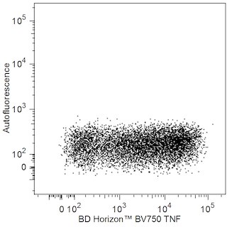
Flow cytometric analysis of TNF expression in stimulated human peripheral blood lymphocytes. Human peripheral blood mononuclear cells were stimulated for 6 hours with Phorbol 12-Myristate 13-Acetate Sigma P-8139; 50 ng/ml final concentration and Ionomycin Sigma I-0634; 1 mug/ml final concentration in the presence of BD GolgiStop Protein Transport Inhibitor containing Monensin Cat. No. 554724 . The cells were harvested, washed with BD Pharmingen Stain Buffer FBS Cat. No. 554656 , and fixed with BD Cytofix Fixation Buffer Cat. No. 554655 . The cells were then washed and stained in BD Perm/Wash Buffer Cat. No. 554723 with either BD Horizon BV750 Mouse IgG1 kappa Isotype Control Cat. No. 566360; Left Plot or BD Horizon BV750 Mouse Anti-Human TNF antibody Cat. No. 566359; Right Plot at 0.5 mug/test. Two-color flow cytometric dot plots showing the correlated expression of TNF or Ig Isotype control staining versus Autofluorescence were derived from gated events with the forward and side light-scatter characteristics of intact lymphocytes. Flow cytometric analysis was performed using a BD LSRFortessa X-20 Cell Analyzer System. Data shown on this Technical Data Sheet are not lot specific. Flow cytometric analysis of TNF expression in stimulated human peripheral blood lymphocytes. Human peripheral blood mononuclear cells were stimulated for 6 hours with Phorbol 12-Myristate 13-Acetate Sigma P-8139; 50 ng/ml final concentration and Ionomycin Sigma I-0634; 1 mug/ml final concentration in the presence of BD GolgiStop Protein Transport Inhibitor containing Monensin Cat. No. 554724 . The cells were harvested, washed with BD Pharmingen Stain Buffer FBS Cat. No. 554656 , and fixed with BD Cytofix Fixation Buffer Cat. No. 554655 . The cells were then washed and stained in BD Perm/Wash Buffer Cat. No. 554723 with either BD Horizon BV750 Mouse IgG1 kappa Isotype Control Cat. No. 566360; Left Plot or BD Horizon BV750 Mouse Anti-Human TNF antibody Cat. No. 566359; Right Plot at 0.5 mug/test. Two-color flow cytometric dot plots showing the correlated expression of TNF or Ig Isotype control staining versus Autofluorescence were derived from gated events with the forward and side light-scatter characteristics of intact lymphocytes. Flow cytometric analysis was performed using a BD LSRFortessa X-20 Cell Analyzer System. Data shown on this Technical Data Sheet are not lot specific. Flow cytometric analysis of TNF expression in stimulated human peripheral blood lymphocytes. Human peripheral blood mononuclear cells were stimulated for 6 hours with Phorbol 12-Myristate 13-Acetate Sigma P-8139; 50 ng/ml final concentration and Ionomycin Sigma I-0634; 1 mug/ml final concentration in the presence of BD GolgiStop Protein Transport Inhibitor containing Monensin Cat. No. 554724 . The cells were harvested, washed with BD Pharmingen Stain Buffer FBS Cat. No. 554656 , and fixed with BD Cytofix Fixation Buffer Cat. No. 554655 . The cells were then washed and stained in BD Perm/Wash Buffer Cat. No. 554723 with either BD Horizon BV750 Mouse IgG1 kappa Isotype Control Cat. No. 566360; Left Plot or BD Horizon BV750 Mouse Anti-Human TNF antibody Cat. No. 566359; Right Plot at 0.5 mug/test. Two-color flow cytometric dot plots showing the correlated expression of TNF or Ig Isotype control staining versus Autofluorescence were derived from gated events with the forward and side light-scatter characteristics of intact lymphocytes. Flow cytometric analysis was performed using a BD LSRFortessa X-20 Cell Analyzer System. Data shown on this Technical Data Sheet are not lot specific. Show More Flow cytometric analysis of TNF expression in stimulated human peripheral blood lymphocytes. Human peripheral blood mononuclear cells were stimulated for 6 hours with Phorbol 12-Myristate 13-Acetate Sigma P-8139; 50 ng/ml final concentration and Ionomycin Sigma I-0634; 1 mug/ml final concentration in the presence of BD GolgiStop Protein Transport Inhibitor containing Monensin Cat. No. 554724 . The cells were harvested, washed with BD Pharmingen Stain Buffer FBS Cat. No. 554656 , and fixed with BD Cytofix Fixation Buffer Cat. No. 554655 . The cells were then washed and stained in BD Perm/Wash Buffer Cat. No. 554723 with either BD Horizon BV750 Mouse IgG1 kappa Isotype Control Cat. No. 566360; Left Plot or BD Horizon BV750 Mouse Anti-Human TNF antibody Cat. No. 566359; Right Plot at 0.5 mug/test. Two-color flow cytometric dot plots showing the correlated expression of TNF or Ig Isotype control staining versus Autofluorescence were derived from gated events with the forward and side light-scatter characteristics of intact lymphocytes. Flow cytometric analysis was performed using a BD LSRFortessa X-20 Cell Analyzer System. Data shown on this Technical Data Sheet are not lot specific. Show More Flow cytometric analysis of TNF expression in stimulated human peripheral blood lymphocytes. Human peripheral blood mononuclear cells were stimulated for 6 hours with Phorbol 12-Myristate 13-Acetate Sigma P-8139; 50 ng/ml final concentration and Ionomycin Sigma I-0634; 1 mug/ml final concentration in the presence of BD GolgiStop Protein Transport Inhibitor containing Monensin Cat. No. 554724 . The cells were harvested, washed with BD Pharmingen Stain Buffer FBS Cat. No. 554656 , and fixed with BD Cytofix Fixation Buffer Cat. No. 554655 . The cells were then washed and stained in BD Perm/Wash Buffer Cat. No. 554723 with either BD Horizon BV750 Mouse IgG1 kappa Isotype Control Cat. No. 566360; Left Plot or BD Horizon BV750 Mouse Anti-Human TNF antibody Cat. No. 566359; Right Plot at 0.5 mug/test. Two-color flow cytometric dot plots showing the correlated expression of TNF or Ig Isotype control staining versus Autofluorescence were derived from gated events with the forward and side light-scatter characteristics of intact lymphocytes. Flow cytometric analysis was performed using a BD LSRFortessa X-20 Cell Analyzer System. Data shown on this Technical Data Sheet are not lot specific. Flow cytometric analysis of TNF expression in stimulated human peripheral blood lymphocytes. Human peripheral blood mononuclear cells were stimulated for 6 hours with Phorbol 12-Myristate 13-Acetate Sigma P-8139; 50 ng/ml final concentration and Ionomycin Sigma I-0634; 1 mug/ml final concentration in the presence of BD GolgiStop Protein Transport Inhibitor containing Monensin Cat. No. 554724 . The cells were harvested, washed with BD Pharmingen Stain Buffer FBS Cat. No. 554656 , and fixed with BD Cytofix Fixation Buffer Cat. No. 554655 . The cells were then washed and stained in BD Perm/Wash Buffer Cat. No. 554723 with either BD Horizon BV750 Mouse IgG1 kappa Isotype Control Cat. No. 566360; Left Plot or BD Horizon BV750 Mouse Anti-Human TNF antibody Cat. No. 566359; Right Plot at 0.5 mug/test. Two-color flow cytometric dot plots showing the correlated expression of TNF or Ig Isotype control staining versus Autofluorescence were derived from gated events with the forward and side light-scatter characteristics of intact lymphocytes. Flow cytometric analysis was performed using a BD LSRFortessa X-20 Cell Analyzer System. Data shown on this Technical Data Sheet are not lot specific.