
BD PharmingenTM Alexa FluorTM 488 Mouse Anti-C-Peptide
571064 · 25 ug
BDB Bioscience · Cat: 571064
Reach 3+ verified distributors
Manufacturer product page
bdbiosciences.com/en-us/products/reagents/flow-cytometry-rea...Description
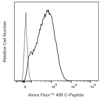
Flow cytometric analysis of C-Peptide expression in Human Insulin-transfected 293F cells and a Mouse insulinoma cell line. Left Plot - Human 293F untransfected dashed line histogram and Insulin-transfected solid line histogram cells were fixed with BD Cytofix Fixation Buffer Cat. No. 554655 and permeabilized with BD Phosflow Perm Buffer III Cat. No. 558050 . The cells were then washed and stained with Alexa Fluor 488 Mouse Anti-C-Peptide antibody Cat. No. 570993/571064; solid line histogram at 0.5 mug/test. Right Plot - Cells from the Mouse Beta-TC-6 Insulinoma, ATCC CRL-3605 cell line were fixed with BD Cytofix Fixation Buffer and permeabilized with BD Phosflow Perm Buffer III. The cells were washed and stained with either Alexa Fluor 488 Mouse IgG1, kappa Isotype Control Cat. No. 565572; dashed line histogram or Alexa Fluor 488 Mouse Anti-C-Peptide antibody solid line histogram at 0.5 mug/test. The fluorescence histograms showing C-Peptide expression were derived from gated events with the forward and side light-scatter characteristics of intact cells. Flow cytometry and data analysis were performed using a BD FACSCanto II Flow Cytometry System and FlowJo Software. Data shown on this Technical Data Sheet is not lot specific. Flow cytometric analysis of C-Peptide expression in Human Insulin-transfected 293F cells and a Mouse insulinoma cell line. Left Plot - Human 293F untransfected dashed line histogram and Insulin-transfected solid line histogram cells were fixed with BD Cytofix Fixation Buffer Cat. No. 554655 and permeabilized with BD Phosflow Perm Buffer III Cat. No. 558050 . The cells were then washed and stained with Alexa Fluor 488 Mouse Anti-C-Peptide antibody Cat. No. 570993/571064; solid line histogram at 0.5 mug/test. Right Plot - Cells from the Mouse Beta-TC-6 Insulinoma, ATCC CRL-3605 cell line were fixed with BD Cytofix Fixation Buffer and permeabilized with BD Phosflow Perm Buffer III. The cells were washed and stained with either Alexa Fluor 488 Mouse IgG1, kappa Isotype Control Cat. No. 565572; dashed line histogram or Alexa Fluor 488 Mouse Anti-C-Peptide antibody solid line histogram at 0.5 mug/test. The fluorescence histograms showing C-Peptide expression were derived from gated events with the forward and side light-scatter characteristics of intact cells. Flow cytometry and data analysis were performed using a BD FACSCanto II Flow Cytometry System and FlowJo Software. Data shown on this Technical Data Sheet is not lot specific. Show More Flow cytometric analysis of C-Peptide expression in Human Insulin-transfected 293F cells and a Mouse insulinoma cell line. Left Plot - Human 293F untransfected dashed line histogram and Insulin-transfected solid line histogram cells were fixed with BD Cytofix Fixation Buffer Cat. No. 554655 and permeabilized with BD Phosflow Perm Buffer III Cat. No. 558050 . The cells were then washed and stained with Alexa Fluor 488 Mouse Anti-C-Peptide antibody Cat. No. 570993/571064; solid line histogram at 0.5 mug/test. Right Plot - Cells from the Mouse Beta-TC-6 Insulinoma, ATCC CRL-3605 cell line were fixed with BD Cytofix Fixation Buffer and permeabilized with BD Phosflow Perm Buffer III. The cells were washed and stained with either Alexa Fluor 488 Mouse IgG1, kappa Isotype Control Cat. No. 565572; dashed line histogram or Alexa Fluor 488 Mouse Anti-C-Peptide antibody solid line histogram at 0.5 mug/test. The fluorescence histograms showing C-Peptide expression were derived from gated events with the forward and side light-scatter characteristics of intact cells. Flow cytometry and data analysis were performed using a BD FACSCanto II Flow Cytometry System and FlowJo Software. Data shown on this Technical Data Sheet is not lot specific.