
BD PharmingenTM PE Mouse Anti-Human AXL
572242 · 100 Tests
BDB Bioscience · Cat: 572242
Reach 3+ verified distributors
Manufacturer product page
bdbiosciences.com/en-us/products/reagents/flow-cytometry-rea...Description
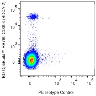
Multicolor flow cytometric analysis of AXL expression on Human A431 cells or Human peripheral blood mononuclear cells. Left Plot: Cells from the Human A431 Epidermoid Carcinoma, ATCC CRL-1555 cell line were preincubated with Human BD Fc Block Cat. No. 564220 and stained with either PE Mouse IgG1, kappa Isotype Control Cat. No. 554680; dashed line histogram or PE Mouse Anti-Human AXL antibody Cat. No. 572242/572243; solid line histogram . DAPI Solution Cat. No. 564907 was added to cells right before analysis. The fluorescence histograms showing AXL expression or Ig Isotype control staining were derived from gated events with the forward and side light-scatter characteristics of viable DAPI-negative cells. Middle and Right Plots: Freshly isolated Human peripheral blood mononuclear cells were preincubated with Human BD Fc Block. The cells were then stained with APC Mouse Anti-Human CD3 Cat. No. 561811 , APC Mouse Anti-Human CD19 Cat. No. 555415 , APC Mouse Anti-Human CD14 Cat. No. 555399 , APC Mouse Anti-Human CD15 Cat. No. 551376 , BD OptiBuild RB780 Mouse Anti-Human CD303 BDCA-2 Cat. No. 755529 and with either PE Mouse IgG1, kappa Isotype Control Middle Plot or PE Mouse Anti-AXL antibody Right Plot . DAPI Solution was added to cells right before analysis. The bivariate pseudocolor density plot showing the correlated expression of AXL or Ig Isotype control staining versus CD303 BDCA-2 was derived from CD3-CD19-CD14-CD15- gated events with the forward and side light-scatter characteristics of viable DAPI-negative cells. Flow cytometry and data analysis were performed using a BD LSRFortessa Cell Analyzer System and FlowJo Software. The above is qualification data only and does not represent a specific OptiBuild lot. Multicolor flow cytometric analysis of AXL expression on Human A431 cells or Human peripheral blood mononuclear cells. Left Plot: Cells from the Human A431 Epidermoid Carcinoma, ATCC CRL-1555 cell line were preincubated with Human BD Fc Block Cat. No. 564220 and stained with either PE Mouse IgG1, kappa Isotype Control Cat. No. 554680; dashed line histogram or PE Mouse Anti-Human AXL antibody Cat. No. 572242/572243; solid line histogram . DAPI Solution Cat. No. 564907 was added to cells right before analysis. The fluorescence histograms showing AXL expression or Ig Isotype control staining were derived from gated events with the forward and side light-scatter characteristics of viable DAPI-negative cells. Middle and Right Plots: Freshly isolated Human peripheral blood mononuclear cells were preincubated with Human BD Fc Block. The cells were then stained with APC Mouse Anti-Human CD3 Cat. No. 561811 , APC Mouse Anti-Human CD19 Cat. No. 555415 , APC Mouse Anti-Human CD14 Cat. No. 555399 , APC Mouse Anti-Human CD15 Cat. No. 551376 , BD OptiBuild RB780 Mouse Anti-Human CD303 BDCA-2 Cat. No. 755529 and with either PE Mouse IgG1, kappa Isotype Control Middle Plot or PE Mouse Anti-AXL antibody Right Plot . DAPI Solution was added to cells right before analysis. The bivariate pseudocolor density plot showing the correlated expression of AXL or Ig Isotype control staining versus CD303 BDCA-2 was derived from CD3-CD19-CD14-CD15- gated events with the forward and side light-scatter characteristics of viable DAPI-negative cells. Flow cytometry and data analysis were performed using a BD LSRFortessa Cell Analyzer System and FlowJo Software. The above is qualification data only and does not represent a specific OptiBuild lot. Multicolor flow cytometric analysis of AXL expression on Human A431 cells or Human peripheral blood mononuclear cells. Left Plot: Cells from the Human A431 Epidermoid Carcinoma, ATCC CRL-1555 cell line were preincubated with Human BD Fc Block Cat. No. 564220 and stained with either PE Mouse IgG1, kappa Isotype Control Cat. No. 554680; dashed line histogram or PE Mouse Anti-Human AXL antibody Cat. No. 572242/572243; solid line histogram . DAPI Solution Cat. No. 564907 was added to cells right before analysis. The fluorescence histograms showing AXL expression or Ig Isotype control staining were derived from gated events with the forward and side light-scatter characteristics of viable DAPI-negative cells. Middle and Right Plots: Freshly isolated Human peripheral blood mononuclear cells were preincubated with Human BD Fc Block. The cells were then stained with APC Mouse Anti-Human CD3 Cat. No. 561811 , APC Mouse Anti-Human CD19 Cat. No. 555415 , APC Mouse Anti-Human CD14 Cat. No. 555399 , APC Mouse Anti-Human CD15 Cat. No. 551376 , BD OptiBuild RB780 Mouse Anti-Human CD303 BDCA-2 Cat. No. 755529 and with either PE Mouse IgG1, kappa Isotype Control Middle Plot or PE Mouse Anti-AXL antibody Right Plot . DAPI Solution was added to cells right before analysis. The bivariate pseudocolor density plot showing the correlated expression of AXL or Ig Isotype control staining versus CD303 BDCA-2 was derived from CD3-CD19-CD14-CD15- gated events with the forward and side light-scatter characteristics of viable DAPI-negative cells. Flow cytometry and data analysis were performed using a BD LSRFortessa Cell Analyzer System and FlowJo Software. The above is qualification data only and does not represent a specific OptiBuild lot. Show More Multicolor flow cytometric analysis of AXL expression on Human A431 cells or Human peripheral blood mononuclear cells. Left Plot: Cells from the Human A431 Epidermoid Carcinoma, ATCC CRL-1555 cell line were preincubated with Human BD Fc Block Cat. No. 564220 and stained with either PE Mouse IgG1, kappa Isotype Control Cat. No. 554680; dashed line histogram or PE Mouse Anti-Human AXL antibody Cat. No. 572242/572243; solid line histogram . DAPI Solution Cat. No. 564907 was added to cells right before analysis. The fluorescence histograms showing AXL expression or Ig Isotype control staining were derived from gated events with the forward and side light-scatter characteristics of viable DAPI-negative cells. Middle and Right Plots: Freshly isolated Human peripheral blood mononuclear cells were preincubated with Human BD Fc Block. The cells were then stained with APC Mouse Anti-Human CD3 Cat. No. 561811 , APC Mouse Anti-Human CD19 Cat. No. 555415 , APC Mouse Anti-Human CD14 Cat. No. 555399 , APC Mouse Anti-Human CD15 Cat. No. 551376 , BD OptiBuild RB780 Mouse Anti-Human CD303 BDCA-2 Cat. No. 755529 and with either PE Mouse IgG1, kappa Isotype Control Middle Plot or PE Mouse Anti-AXL antibody Right Plot . DAPI Solution was added to cells right before analysis. The bivariate pseudocolor density plot showing the correlated expression of AXL or Ig Isotype control staining versus CD303 BDCA-2 was derived from CD3-CD19-CD14-CD15- gated events with the forward and side light-scatter characteristics of viable DAPI-negative cells. Flow cytometry and data analysis were performed using a BD LSRFortessa Cell Analyzer System and FlowJo Software. The above is qualification data only and does not represent a specific OptiBuild lot. Show More Multicolor flow cytometric analysis of AXL expression on Human A431 cells or Human peripheral blood mononuclear cells. Left Plot: Cells from the Human A431 Epidermoid Carcinoma, ATCC CRL-1555 cell line were preincubated with Human BD Fc Block Cat. No. 564220 and stained with either PE Mouse IgG1, kappa Isotype Control Cat. No. 554680; dashed line histogram or PE Mouse Anti-Human AXL antibody Cat. No. 572242/572243; solid line histogram . DAPI Solution Cat. No. 564907 was added to cells right before analysis. The fluorescence histograms showing AXL expression or Ig Isotype control staining were derived from gated events with the forward and side light-scatter characteristics of viable DAPI-negative cells. Middle and Right Plots: Freshly isolated Human peripheral blood mononuclear cells were preincubated with Human BD Fc Block. The cells were then stained with APC Mouse Anti-Human CD3 Cat. No. 561811 , APC Mouse Anti-Human CD19 Cat. No. 555415 , APC Mouse Anti-Human CD14 Cat. No. 555399 , APC Mouse Anti-Human CD15 Cat. No. 551376 , BD OptiBuild RB780 Mouse Anti-Human CD303 BDCA-2 Cat. No. 755529 and with either PE Mouse IgG1, kappa Isotype Control Middle Plot or PE Mouse Anti-AXL antibody Right Plot . DAPI Solution was added to cells right before analysis. The bivariate pseudocolor density plot showing the correlated expression of AXL or Ig Isotype control staining versus CD303 BDCA-2 was derived from CD3-CD19-CD14-CD15- gated events with the forward and side light-scatter characteristics of viable DAPI-negative cells. Flow cytometry and data analysis were performed using a BD LSRFortessa Cell Analyzer System and FlowJo Software. The above is qualification data only and does not represent a specific OptiBuild lot. Multicolor flow cytometric analysis of AXL expression on Human A431 cells or Human peripheral blood mononuclear cells. Left Plot: Cells from the Human A431 Epidermoid Carcinoma, ATCC CRL-1555 cell line were preincubated with Human BD Fc Block Cat. No. 564220 and stained with either PE Mouse IgG1, kappa Isotype Control Cat. No. 554680; dashed line histogram or PE Mouse Anti-Human AXL antibody Cat. No. 572242/572243; solid line histogram . DAPI Solution Cat. No. 564907 was added to cells right before analysis. The fluorescence histograms showing AXL expression or Ig Isotype control staining were derived from gated events with the forward and side light-scatter characteristics of viable DAPI-negative cells. Middle and Right Plots: Freshly isolated Human peripheral blood mononuclear cells were preincubated with Human BD Fc Block. The cells were then stained with APC Mouse Anti-Human CD3 Cat. No. 561811 , APC Mouse Anti-Human CD19 Cat. No. 555415 , APC Mouse Anti-Human CD14 Cat. No. 555399 , APC Mouse Anti-Human CD15 Cat. No. 551376 , BD OptiBuild RB780 Mouse Anti-Human CD303 BDCA-2 Cat. No. 755529 and with either PE Mouse IgG1, kappa Isotype Control Middle Plot or PE Mouse Anti-AXL antibody Right Plot . DAPI Solution was added to cells right before analysis. The bivariate pseudocolor density plot showing the correlated expression of AXL or Ig Isotype control staining versus CD303 BDCA-2 was derived from CD3-CD19-CD14-CD15- gated events with the forward and side light-scatter characteristics of viable DAPI-negative cells. Flow cytometry and data analysis were performed using a BD LSRFortessa Cell Analyzer System and FlowJo Software. The above is qualification data only and does not represent a specific OptiBuild lot.