
BD PharmingenTM PE Mouse Anti-Human Langerin (CD207)
564727 ·
BDB Bioscience · Cat: 564727
Reach 3+ verified distributors
Manufacturer product page
bdbiosciences.com/en-us/products/reagents/flow-cytometry-rea...Description
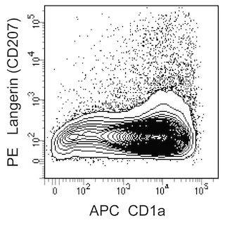
Multicolor flow cytometric analysis of human Langerin CD207 expression by monocyte-derived Langerhans cells. Peripheral blood monocytes were cultured with Recombinant Human GM-CSF Cat. No. 550068; 100 ng/ml , IL-4 Cat. No. 554605; 20 ng/ml , and TGF-beta Corning; Cat. No. 354039; 40 ng/ml proteins for 3 days followed by an additional 3-day culture with GM-CSF and TGF-beta. The cells were washed and stained with APC Mouse Anti-Human CD1a antibody Cat. No. 559775/561755 and BD Horizon Fixable Viability Stain 450 Cat. No. 562247 . After washing, the cells were fixed and permeabilized with BD Cytofix/Cytoperm Fixation and Permeabilization Solution Cat. No. 554722 . The cells were subsequently washed and stained in BD Perm/Wash Buffer Cat. No. 554723 with either PE Mouse IgG1, kappa Isotype Control Cat. No. 554680; Left Panel or PE Mouse Anti-Human Langerin CD207 antibody Cat. No. 564727; Right Panel . Flow cytometric contour plots showing the correlated expression of Langerin CD207 or Ig Isotype control staining versus CD1a were derived from gated events with the forward and side light-scatter, and viability stain characteristics of intact cells. Flow cytometric analysis was performed using a BD LSRFortessa X-20 Cell Analyzer System. Multicolor flow cytometric analysis of human Langerin CD207 expression by monocyte-derived Langerhans cells. Peripheral blood monocytes were cultured with Recombinant Human GM-CSF Cat. No. 550068; 100 ng/ml , IL-4 Cat. No. 554605; 20 ng/ml , and TGF-beta Corning; Cat. No. 354039; 40 ng/ml proteins for 3 days followed by an additional 3-day culture with GM-CSF and TGF-beta. The cells were washed and stained with APC Mouse Anti-Human CD1a antibody Cat. No. 559775/561755 and BD Horizon Fixable Viability Stain 450 Cat. No. 562247 . After washing, the cells were fixed and permeabilized with BD Cytofix/Cytoperm Fixation and Permeabilization Solution Cat. No. 554722 . The cells were subsequently washed and stained in BD Perm/Wash Buffer Cat. No. 554723 with either PE Mouse IgG1, kappa Isotype Control Cat. No. 554680; Left Panel or PE Mouse Anti-Human Langerin CD207 antibody Cat. No. 564727; Right Panel . Flow cytometric contour plots showing the correlated expression of Langerin CD207 or Ig Isotype control staining versus CD1a were derived from gated events with the forward and side light-scatter, and viability stain characteristics of intact cells. Flow cytometric analysis was performed using a BD LSRFortessa X-20 Cell Analyzer System. Multicolor flow cytometric analysis of human Langerin CD207 expression by monocyte-derived Langerhans cells. Peripheral blood monocytes were cultured with Recombinant Human GM-CSF Cat. No. 550068; 100 ng/ml , IL-4 Cat. No. 554605; 20 ng/ml , and TGF-beta Corning; Cat. No. 354039; 40 ng/ml proteins for 3 days followed by an additional 3-day culture with GM-CSF and TGF-beta. The cells were washed and stained with APC Mouse Anti-Human CD1a antibody Cat. No. 559775/561755 and BD Horizon Fixable Viability Stain 450 Cat. No. 562247 . After washing, the cells were fixed and permeabilized with BD Cytofix/Cytoperm Fixation and Permeabilization Solution Cat. No. 554722 . The cells were subsequently washed and stained in BD Perm/Wash Buffer Cat. No. 554723 with either PE Mouse IgG1, kappa Isotype Control Cat. No. 554680; Left Panel or PE Mouse Anti-Human Langerin CD207 antibody Cat. No. 564727; Right Panel . Flow cytometric contour plots showing the correlated expression of Langerin CD207 or Ig Isotype control staining versus CD1a were derived from gated events with the forward and side light-scatter, and viability stain characteristics of intact cells. Flow cytometric analysis was performed using a BD LSRFortessa X-20 Cell Analyzer System. Show More Multicolor flow cytometric analysis of human Langerin CD207 expression by monocyte-derived Langerhans cells. Peripheral blood monocytes were cultured with Recombinant Human GM-CSF Cat. No. 550068; 100 ng/ml , IL-4 Cat. No. 554605; 20 ng/ml , and TGF-beta Corning; Cat. No. 354039; 40 ng/ml proteins for 3 days followed by an additional 3-day culture with GM-CSF and TGF-beta. The cells were washed and stained with APC Mouse Anti-Human CD1a antibody Cat. No. 559775/561755 and BD Horizon Fixable Viability Stain 450 Cat. No. 562247 . After washing, the cells were fixed and permeabilized with BD Cytofix/Cytoperm Fixation and Permeabilization Solution Cat. No. 554722 . The cells were subsequently washed and stained in BD Perm/Wash Buffer Cat. No. 554723 with either PE Mouse IgG1, kappa Isotype Control Cat. No. 554680; Left Panel or PE Mouse Anti-Human Langerin CD207 antibody Cat. No. 564727; Right Panel . Flow cytometric contour plots showing the correlated expression of Langerin CD207 or Ig Isotype control staining versus CD1a were derived from gated events with the forward and side light-scatter, and viability stain characteristics of intact cells. Flow cytometric analysis was performed using a BD LSRFortessa X-20 Cell Analyzer System. Show More Multicolor flow cytometric analysis of human Langerin CD207 expression by monocyte-derived Langerhans cells. Peripheral blood monocytes were cultured with Recombinant Human GM-CSF Cat. No. 550068; 100 ng/ml , IL-4 Cat. No. 554605; 20 ng/ml , and TGF-beta Corning; Cat. No. 354039; 40 ng/ml proteins for 3 days followed by an additional 3-day culture with GM-CSF and TGF-beta. The cells were washed and stained with APC Mouse Anti-Human CD1a antibody Cat. No. 559775/561755 and BD Horizon Fixable Viability Stain 450 Cat. No. 562247 . After washing, the cells were fixed and permeabilized with BD Cytofix/Cytoperm Fixation and Permeabilization Solution Cat. No. 554722 . The cells were subsequently washed and stained in BD Perm/Wash Buffer Cat. No. 554723 with either PE Mouse IgG1, kappa Isotype Control Cat. No. 554680; Left Panel or PE Mouse Anti-Human Langerin CD207 antibody Cat. No. 564727; Right Panel . Flow cytometric contour plots showing the correlated expression of Langerin CD207 or Ig Isotype control staining versus CD1a were derived from gated events with the forward and side light-scatter, and viability stain characteristics of intact cells. Flow cytometric analysis was performed using a BD LSRFortessa X-20 Cell Analyzer System. Multicolor flow cytometric analysis of human Langerin CD207 expression by monocyte-derived Langerhans cells. Peripheral blood monocytes were cultured with Recombinant Human GM-CSF Cat. No. 550068; 100 ng/ml , IL-4 Cat. No. 554605; 20 ng/ml , and TGF-beta Corning; Cat. No. 354039; 40 ng/ml proteins for 3 days followed by an additional 3-day culture with GM-CSF and TGF-beta. The cells were washed and stained with APC Mouse Anti-Human CD1a antibody Cat. No. 559775/561755 and BD Horizon Fixable Viability Stain 450 Cat. No. 562247 . After washing, the cells were fixed and permeabilized with BD Cytofix/Cytoperm Fixation and Permeabilization Solution Cat. No. 554722 . The cells were subsequently washed and stained in BD Perm/Wash Buffer Cat. No. 554723 with either PE Mouse IgG1, kappa Isotype Control Cat. No. 554680; Left Panel or PE Mouse Anti-Human Langerin CD207 antibody Cat. No. 564727; Right Panel . Flow cytometric contour plots showing the correlated expression of Langerin CD207 or Ig Isotype control staining versus CD1a were derived from gated events with the forward and side light-scatter, and viability stain characteristics of intact cells. Flow cytometric analysis was performed using a BD LSRFortessa X-20 Cell Analyzer System.