
BD PharmingenTM PE Rat Anti-Mouse CD326
563477 ·
BDB Bioscience · Cat: 563477
Reach 3+ verified distributors
Manufacturer product page
bdbiosciences.com/en-us/products/reagents/flow-cytometry-rea...
Description
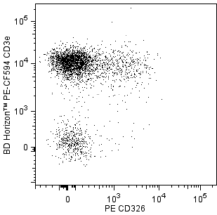
Two-color analysis of CD326 expression on mouse thymocytes and splenic T lymphocytes. BALB/c mouse thymocytes and splenic leucocytes were preincubated with Purified Rat Anti-Mouse CD16/CD32 antibody Mouse BD Fc Block Cat. No. 553141/553142 and stained with PE Rat IgG2a, kappa Isotype Control Cat. No. 554689 or PE Rat Anti-Mouse CD326 antibody Cat. No. 563477 . Left Panel: Thymocytes were further stained with FITC Rat Anti-Mouse CD4 553047/553046/561835 and APC Rat Anti-Mouse CD8a 553035/561093 antibodies. The CD326 solid line histogram and Ig Isotype Control dashed line histogram fluorescence histograms were derived from CD4- and CD8-negative gated events with the forward and side light-scatter characteristics of viable thymocytes. Middle and Right Panels: The splenic leucocytes were further stained with BD Horizon PE-CF594 Hamster Anti-Mouse CD3e Cat. No 562332/562286 and APC Rat Anti-Mouse CD25 Cat. No. 557192/561048 antibodies. Two-color flow cytometric dot plots show the correlated expression patterns of Ig Isotype control staining Middle Panel or CD326 Right Panel versus CD3e for CD25+ gated events with the forward and side light-scatter characteristics of viable splenic leucocytes. A small population of CD3+CD25+CD326+ cells were detected Right Panel , whereas the CD25- T cells do not express detectable levels of CD326 data not shown . Flow cytometric analysis was performed using a BD LSR II Flow Cytometer System. Two-color analysis of CD326 expression on mouse thymocytes and splenic T lymphocytes. BALB/c mouse thymocytes and splenic leucocytes were preincubated with Purified Rat Anti-Mouse CD16/CD32 antibody Mouse BD Fc Block Cat. No. 553141/553142 and stained with PE Rat IgG2a, kappa Isotype Control Cat. No. 554689 or PE Rat Anti-Mouse CD326 antibody Cat. No. 563477 . Left Panel: Thymocytes were further stained with FITC Rat Anti-Mouse CD4 553047/553046/561835 and APC Rat Anti-Mouse CD8a 553035/561093 antibodies. The CD326 solid line histogram and Ig Isotype Control dashed line histogram fluorescence histograms were derived from CD4- and CD8-negative gated events with the forward and side light-scatter characteristics of viable thymocytes. Middle and Right Panels: The splenic leucocytes were further stained with BD Horizon PE-CF594 Hamster Anti-Mouse CD3e Cat. No 562332/562286 and APC Rat Anti-Mouse CD25 Cat. No. 557192/561048 antibodies. Two-color flow cytometric dot plots show the correlated expression patterns of Ig Isotype control staining Middle Panel or CD326 Right Panel versus CD3e for CD25+ gated events with the forward and side light-scatter characteristics of viable splenic leucocytes. A small population of CD3+CD25+CD326+ cells were detected Right Panel , whereas the CD25- T cells do not express detectable levels of CD326 data not shown . Flow cytometric analysis was performed using a BD LSR II Flow Cytometer System. Two-color analysis of CD326 expression on mouse thymocytes and splenic T lymphocytes. BALB/c mouse thymocytes and splenic leucocytes were preincubated with Purified Rat Anti-Mouse CD16/CD32 antibody Mouse BD Fc Block Cat. No. 553141/553142 and stained with PE Rat IgG2a, kappa Isotype Control Cat. No. 554689 or PE Rat Anti-Mouse CD326 antibody Cat. No. 563477 . Left Panel: Thymocytes were further stained with FITC Rat Anti-Mouse CD4 553047/553046/561835 and APC Rat Anti-Mouse CD8a 553035/561093 antibodies. The CD326 solid line histogram and Ig Isotype Control dashed line histogram fluorescence histograms were derived from CD4- and CD8-negative gated events with the forward and side light-scatter characteristics of viable thymocytes. Middle and Right Panels: The splenic leucocytes were further stained with BD Horizon PE-CF594 Hamster Anti-Mouse CD3e Cat. No 562332/562286 and APC Rat Anti-Mouse CD25 Cat. No. 557192/561048 antibodies. Two-color flow cytometric dot plots show the correlated expression patterns of Ig Isotype control staining Middle Panel or CD326 Right Panel versus CD3e for CD25+ gated events with the forward and side light-scatter characteristics of viable splenic leucocytes. A small population of CD3+CD25+CD326+ cells were detected Right Panel , whereas the CD25- T cells do not express detectable levels of CD326 data not shown . Flow cytometric analysis was performed using a BD LSR II Flow Cytometer System. Show More Two-color analysis of CD326 expression on mouse thymocytes and splenic T lymphocytes. BALB/c mouse thymocytes and splenic leucocytes were preincubated with Purified Rat Anti-Mouse CD16/CD32 antibody Mouse BD Fc Block Cat. No. 553141/553142 and stained with PE Rat IgG2a, kappa Isotype Control Cat. No. 554689 or PE Rat Anti-Mouse CD326 antibody Cat. No. 563477 . Left Panel: Thymocytes were further stained with FITC Rat Anti-Mouse CD4 553047/553046/561835 and APC Rat Anti-Mouse CD8a 553035/561093 antibodies. The CD326 solid line histogram and Ig Isotype Control dashed line histogram fluorescence histograms were derived from CD4- and CD8-negative gated events with the forward and side light-scatter characteristics of viable thymocytes. Middle and Right Panels: The splenic leucocytes were further stained with BD Horizon PE-CF594 Hamster Anti-Mouse CD3e Cat. No 562332/562286 and APC Rat Anti-Mouse CD25 Cat. No. 557192/561048 antibodies. Two-color flow cytometric dot plots show the correlated expression patterns of Ig Isotype control staining Middle Panel or CD326 Right Panel versus CD3e for CD25+ gated events with the forward and side light-scatter characteristics of viable splenic leucocytes. A small population of CD3+CD25+CD326+ cells were detected Right Panel , whereas the CD25- T cells do not express detectable levels of CD326 data not shown . Flow cytometric analysis was performed using a BD LSR II Flow Cytometer System. Show More Two-color analysis of CD326 expression on mouse thymocytes and splenic T lymphocytes. BALB/c mouse thymocytes and splenic leucocytes were preincubated with Purified Rat Anti-Mouse CD16/CD32 antibody Mouse BD Fc Block Cat. No. 553141/553142 and stained with PE Rat IgG2a, kappa Isotype Control Cat. No. 554689 or PE Rat Anti-Mouse CD326 antibody Cat. No. 563477 . Left Panel: Thymocytes were further stained with FITC Rat Anti-Mouse CD4 553047/553046/561835 and APC Rat Anti-Mouse CD8a 553035/561093 antibodies. The CD326 solid line histogram and Ig Isotype Control dashed line histogram fluorescence histograms were derived from CD4- and CD8-negative gated events with the forward and side light-scatter characteristics of viable thymocytes. Middle and Right Panels: The splenic leucocytes were further stained with BD Horizon PE-CF594 Hamster Anti-Mouse CD3e Cat. No 562332/562286 and APC Rat Anti-Mouse CD25 Cat. No. 557192/561048 antibodies. Two-color flow cytometric dot plots show the correlated expression patterns of Ig Isotype control staining Middle Panel or CD326 Right Panel versus CD3e for CD25+ gated events with the forward and side light-scatter characteristics of viable splenic leucocytes. A small population of CD3+CD25+CD326+ cells were detected Right Panel , whereas the CD25- T cells do not express detectable levels of CD326 data not shown . Flow cytometric analysis was performed using a BD LSR II Flow Cytometer System. Two-color analysis of CD326 expression on mouse thymocytes and splenic T lymphocytes. BALB/c mouse thymocytes and splenic leucocytes were preincubated with Purified Rat Anti-Mouse CD16/CD32 antibody Mouse BD Fc Block Cat. No. 553141/553142 and stained with PE Rat IgG2a, kappa Isotype Control Cat. No. 554689 or PE Rat Anti-Mouse CD326 antibody Cat. No. 563477 . Left Panel: Thymocytes were further stained with FITC Rat Anti-Mouse CD4 553047/553046/561835 and APC Rat Anti-Mouse CD8a 553035/561093 antibodies. The CD326 solid line histogram and Ig Isotype Control dashed line histogram fluorescence histograms were derived from CD4- and CD8-negative gated events with the forward and side light-scatter characteristics of viable thymocytes. Middle and Right Panels: The splenic leucocytes were further stained with BD Horizon PE-CF594 Hamster Anti-Mouse CD3e Cat. No 562332/562286 and APC Rat Anti-Mouse CD25 Cat. No. 557192/561048 antibodies. Two-color flow cytometric dot plots show the correlated expression patterns of Ig Isotype control staining Middle Panel or CD326 Right Panel versus CD3e for CD25+ gated events with the forward and side light-scatter characteristics of viable splenic leucocytes. A small population of CD3+CD25+CD326+ cells were detected Right Panel , whereas the CD25- T cells do not express detectable levels of CD326 data not shown . Flow cytometric analysis was performed using a BD LSR II Flow Cytometer System.