
BD PharmingenTM PE Rat Anti-Mouse IL-4
554435 · 0.1 mg
BDB Bioscience · Cat: 554435
Reach 3+ verified distributors
Manufacturer product page
bdbiosciences.com/en-us/products/reagents/flow-cytometry-rea...Description
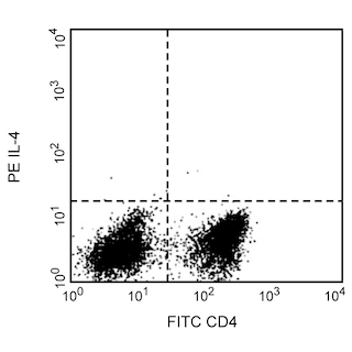
Expression of IL-4 by stimulated CD4+ and CD4-BALB/c spleen cells. BALB/c spleen cells were cultured for 72 h in medium containing Staphylococcus aureus enterotoxin B 2 mug/ml; Sigma, St. Louis, MO , recombinant mouse IL-2 10 U/ml, Cat. No. 550069 and recombinant mouse IL-4 2 ng/ml, Cat. No. 550067 . The cells were harvested and restimulated for 5 h with anti-CD3 145-2C11, Cat. No. 553057 at 2 mug/ml and anti-CD28 clone 37.51, Cat. No. 553294 at 2 mug/ml antibodies in the presence of 3 muM monensin BD GolgiStop, Cat No. 554704 . The splenocytes were then stained with 0.25 mug of FITC-conjugated rat anti-mouse CD4 FITC-RM4-5, Cat. No. 553047 and 0.25 mug of PE-conjugated rat anti-mouse IL-4 antibody PE11B11, Cat. No 554435 by using the Pharmingen staining protocol left panel . To demonstrate staining specificity, the binding of PE-11B11 was blocked by the preincubation of the conjugated antibody with excess recombinant mouse IL-4 0.25 mug; Cat. No. 550067 right panel or by pre-blocking fixed/permeabilized cells with excess purified 11B11 mAb 5.0 mug; Cat. No. 554433 data not shown , prior to staining. An approprate isotype control is PE-conjugated R3-34, Cat. No. 554685 . The quadrant markers for the bivariate dot plots were set based on the autofluorescence control, and verified using the cytokine-blocking or mAb blocking controls. Expression of IL-4 by stimulated CD4+ and CD4-BALB/c spleen cells. BALB/c spleen cells were cultured for 72 h in medium containing Staphylococcus aureus enterotoxin B 2 mug/ml; Sigma, St. Louis, MO , recombinant mouse IL-2 10 U/ml, Cat. No. 550069 and recombinant mouse IL-4 2 ng/ml, Cat. No. 550067 . The cells were harvested and restimulated for 5 h with anti-CD3 145-2C11, Cat. No. 553057 at 2 mug/ml and anti-CD28 clone 37.51, Cat. No. 553294 at 2 mug/ml antibodies in the presence of 3 muM monensin BD GolgiStop, Cat No. 554704 . The splenocytes were then stained with 0.25 mug of FITC-conjugated rat anti-mouse CD4 FITC-RM4-5, Cat. No. 553047 and 0.25 mug of PE-conjugated rat anti-mouse IL-4 antibody PE11B11, Cat. No 554435 by using the Pharmingen staining protocol left panel . To demonstrate staining specificity, the binding of PE-11B11 was blocked by the preincubation of the conjugated antibody with excess recombinant mouse IL-4 0.25 mug; Cat. No. 550067 right panel or by pre-blocking fixed/permeabilized cells with excess purified 11B11 mAb 5.0 mug; Cat. No. 554433 data not shown , prior to staining. An approprate isotype control is PE-conjugated R3-34, Cat. No. 554685 . The quadrant markers for the bivariate dot plots were set based on the autofluorescence control, and verified using the cytokine-blocking or mAb blocking controls. Expression of IL-4 by stimulated CD4+ and CD4-BALB/c spleen cells. BALB/c spleen cells were cultured for 72 h in medium containing Staphylococcus aureus enterotoxin B 2 mug/ml; Sigma, St. Louis, MO , recombinant mouse IL-2 10 U/ml, Cat. No. 550069 and recombinant mouse IL-4 2 ng/ml, Cat. No. 550067 . The cells were harvested and restimulated for 5 h with anti-CD3 145-2C11, Cat. No. 553057 at 2 mug/ml and anti-CD28 clone 37.51, Cat. No. 553294 at 2 mug/ml antibodies in the presence of 3 muM monensin BD GolgiStop, Cat No. 554704 . The splenocytes were then stained with 0.25 mug of FITC-conjugated rat anti-mouse CD4 FITC-RM4-5, Cat. No. 553047 and 0.25 mug of PE-conjugated rat anti-mouse IL-4 antibody PE11B11, Cat. No 554435 by using the Pharmingen staining protocol left panel . To demonstrate staining specificity, the binding of PE-11B11 was blocked by the preincubation of the conjugated antibody with excess recombinant mouse IL-4 0.25 mug; Cat. No. 550067 right panel or by pre-blocking fixed/permeabilized cells with excess purified 11B11 mAb 5.0 mug; Cat. No. 554433 data not shown , prior to staining. An approprate isotype control is PE-conjugated R3-34, Cat. No. 554685 . The quadrant markers for the bivariate dot plots were set based on the autofluorescence control, and verified using the cytokine-blocking or mAb blocking controls. Show More Expression of IL-4 by stimulated CD4+ and CD4-BALB/c spleen cells. BALB/c spleen cells were cultured for 72 h in medium containing Staphylococcus aureus enterotoxin B 2 mug/ml; Sigma, St. Louis, MO , recombinant mouse IL-2 10 U/ml, Cat. No. 550069 and recombinant mouse IL-4 2 ng/ml, Cat. No. 550067 . The cells were harvested and restimulated for 5 h with anti-CD3 145-2C11, Cat. No. 553057 at 2 mug/ml and anti-CD28 clone 37.51, Cat. No. 553294 at 2 mug/ml antibodies in the presence of 3 muM monensin BD GolgiStop, Cat No. 554704 . The splenocytes were then stained with 0.25 mug of FITC-conjugated rat anti-mouse CD4 FITC-RM4-5, Cat. No. 553047 and 0.25 mug of PE-conjugated rat anti-mouse IL-4 antibody PE11B11, Cat. No 554435 by using the Pharmingen staining protocol left panel . To demonstrate staining specificity, the binding of PE-11B11 was blocked by the preincubation of the conjugated antibody with excess recombinant mouse IL-4 0.25 mug; Cat. No. 550067 right panel or by pre-blocking fixed/permeabilized cells with excess purified 11B11 mAb 5.0 mug; Cat. No. 554433 data not shown , prior to staining. An approprate isotype control is PE-conjugated R3-34, Cat. No. 554685 . The quadrant markers for the bivariate dot plots were set based on the autofluorescence control, and verified using the cytokine-blocking or mAb blocking controls. Show More Expression of IL-4 by stimulated CD4+ and CD4-BALB/c spleen cells. BALB/c spleen cells were cultured for 72 h in medium containing Staphylococcus aureus enterotoxin B 2 mug/ml; Sigma, St. Louis, MO , recombinant mouse IL-2 10 U/ml, Cat. No. 550069 and recombinant mouse IL-4 2 ng/ml, Cat. No. 550067 . The cells were harvested and restimulated for 5 h with anti-CD3 145-2C11, Cat. No. 553057 at 2 mug/ml and anti-CD28 clone 37.51, Cat. No. 553294 at 2 mug/ml antibodies in the presence of 3 muM monensin BD GolgiStop, Cat No. 554704 . The splenocytes were then stained with 0.25 mug of FITC-conjugated rat anti-mouse CD4 FITC-RM4-5, Cat. No. 553047 and 0.25 mug of PE-conjugated rat anti-mouse IL-4 antibody PE11B11, Cat. No 554435 by using the Pharmingen staining protocol left panel . To demonstrate staining specificity, the binding of PE-11B11 was blocked by the preincubation of the conjugated antibody with excess recombinant mouse IL-4 0.25 mug; Cat. No. 550067 right panel or by pre-blocking fixed/permeabilized cells with excess purified 11B11 mAb 5.0 mug; Cat. No. 554433 data not shown , prior to staining. An approprate isotype control is PE-conjugated R3-34, Cat. No. 554685 . The quadrant markers for the bivariate dot plots were set based on the autofluorescence control, and verified using the cytokine-blocking or mAb blocking controls. Expression of IL-4 by stimulated CD4+ and CD4-BALB/c spleen cells. BALB/c spleen cells were cultured for 72 h in medium containing Staphylococcus aureus enterotoxin B 2 mug/ml; Sigma, St. Louis, MO , recombinant mouse IL-2 10 U/ml, Cat. No. 550069 and recombinant mouse IL-4 2 ng/ml, Cat. No. 550067 . The cells were harvested and restimulated for 5 h with anti-CD3 145-2C11, Cat. No. 553057 at 2 mug/ml and anti-CD28 clone 37.51, Cat. No. 553294 at 2 mug/ml antibodies in the presence of 3 muM monensin BD GolgiStop, Cat No. 554704 . The splenocytes were then stained with 0.25 mug of FITC-conjugated rat anti-mouse CD4 FITC-RM4-5, Cat. No. 553047 and 0.25 mug of PE-conjugated rat anti-mouse IL-4 antibody PE11B11, Cat. No 554435 by using the Pharmingen staining protocol left panel . To demonstrate staining specificity, the binding of PE-11B11 was blocked by the preincubation of the conjugated antibody with excess recombinant mouse IL-4 0.25 mug; Cat. No. 550067 right panel or by pre-blocking fixed/permeabilized cells with excess purified 11B11 mAb 5.0 mug; Cat. No. 554433 data not shown , prior to staining. An approprate isotype control is PE-conjugated R3-34, Cat. No. 554685 . The quadrant markers for the bivariate dot plots were set based on the autofluorescence control, and verified using the cytokine-blocking or mAb blocking controls.