BD PharmingenTM Purified Mouse anti-MAPKAPK-2 (pT334)
562469 ·
BDB Bioscience · Cat: 562469
Reach 3+ verified distributors
Manufacturer product page
bdbiosciences.com/en-us/products/reagents/flow-cytometry-rea...
Description
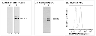
Analyses of MAPKAPK2 pT334 expression. Panel 1: Western blot analysis of MAPKAPK2 pT334 expressed by human THP-1 cells. Lysates 15 mug total cell protein/lane from untreated C and PMA-treated T Phorbol 12-Myristate 13-Acetate, Sigma, Cat. No. P8139; 50 nM, 15 min THP-1 cells were blotted using Purified Mouse Anti-MAPKAPK2 pT334 antibody Cat. No. 562469; 0.5, 0.25, and 0.125 mug/ml for Lanes 1, 2, and 3, respectively , HRP Goat Anti-Mouse Ig Cat. No. 554002 and a chemiluminescent detection system. Panel 2a: Western blot analysis of MAPKAPK2 pT334 expressed by human peripheral blood mononuclear cells PBMC . Lysates from 1 million untreated C and PMA-treated T PBMC were blotted using Purified Mouse Anti-MAPKAPK2 pT334 antibody 2.0 mug/ml as described above. Panel 2b: Flow Cytometric analysis of MAPKAPK2 pT334 expressed by peripheral blood lymphocytes PBL . Human whole blood cells were either not stimulated dashed line histogram or stimulated solid line histogram with 400 nM PMA 15 min, 37C . Cells were fixed in 1X BD Phosflow Lyse/Fix Buffer Cat. No. 558049; 10 min, 37 C and permeabilized in BD Phosflow Perm Buffer III Cat. No. 558050 on ice 30 min . Cells were stained with BD Phosflow PE Mouse Anti-MAPKAPK2 pT334 Cat. No. 562470 antibody. Fluorescence histograms showing MAPKAPK2 pT334 expression were generated for gated events with the forward and side-light scatter characteristics of intact lymphocytes using a BD FACSCanto II Flow Cytometer System. Note: MAPKAPK2 pT334 were identified as 49 kDa bands in the cell lysates by Western blotting. Analyses of MAPKAPK2 pT334 expression. Panel 1: Western blot analysis of MAPKAPK2 pT334 expressed by human THP-1 cells. Lysates 15 mug total cell protein/lane from untreated C and PMA-treated T Phorbol 12-Myristate 13-Acetate, Sigma, Cat. No. P8139; 50 nM, 15 min THP-1 cells were blotted using Purified Mouse Anti-MAPKAPK2 pT334 antibody Cat. No. 562469; 0.5, 0.25, and 0.125 mug/ml for Lanes 1, 2, and 3, respectively , HRP Goat Anti-Mouse Ig Cat. No. 554002 and a chemiluminescent detection system. Panel 2a: Western blot analysis of MAPKAPK2 pT334 expressed by human peripheral blood mononuclear cells PBMC . Lysates from 1 million untreated C and PMA-treated T PBMC were blotted using Purified Mouse Anti-MAPKAPK2 pT334 antibody 2.0 mug/ml as described above. Panel 2b: Flow Cytometric analysis of MAPKAPK2 pT334 expressed by peripheral blood lymphocytes PBL . Human whole blood cells were either not stimulated dashed line histogram or stimulated solid line histogram with 400 nM PMA 15 min, 37C . Cells were fixed in 1X BD Phosflow Lyse/Fix Buffer Cat. No. 558049; 10 min, 37 C and permeabilized in BD Phosflow Perm Buffer III Cat. No. 558050 on ice 30 min . Cells were stained with BD Phosflow PE Mouse Anti-MAPKAPK2 pT334 Cat. No. 562470 antibody. Fluorescence histograms showing MAPKAPK2 pT334 expression were generated for gated events with the forward and side-light scatter characteristics of intact lymphocytes using a BD FACSCanto II Flow Cytometer System. Note: MAPKAPK2 pT334 were identified as 49 kDa bands in the cell lysates by Western blotting. Show More Analyses of MAPKAPK2 pT334 expression. Panel 1: Western blot analysis of MAPKAPK2 pT334 expressed by human THP-1 cells. Lysates 15 mug total cell protein/lane from untreated C and PMA-treated T Phorbol 12-Myristate 13-Acetate, Sigma, Cat. No. P8139; 50 nM, 15 min THP-1 cells were blotted using Purified Mouse Anti-MAPKAPK2 pT334 antibody Cat. No. 562469; 0.5, 0.25, and 0.125 mug/ml for Lanes 1, 2, and 3, respectively , HRP Goat Anti-Mouse Ig Cat. No. 554002 and a chemiluminescent detection system. Panel 2a: Western blot analysis of MAPKAPK2 pT334 expressed by human peripheral blood mononuclear cells PBMC . Lysates from 1 million untreated C and PMA-treated T PBMC were blotted using Purified Mouse Anti-MAPKAPK2 pT334 antibody 2.0 mug/ml as described above. Panel 2b: Flow Cytometric analysis of MAPKAPK2 pT334 expressed by peripheral blood lymphocytes PBL . Human whole blood cells were either not stimulated dashed line histogram or stimulated solid line histogram with 400 nM PMA 15 min, 37C . Cells were fixed in 1X BD Phosflow Lyse/Fix Buffer Cat. No. 558049; 10 min, 37 C and permeabilized in BD Phosflow Perm Buffer III Cat. No. 558050 on ice 30 min . Cells were stained with BD Phosflow PE Mouse Anti-MAPKAPK2 pT334 Cat. No. 562470 antibody. Fluorescence histograms showing MAPKAPK2 pT334 expression were generated for gated events with the forward and side-light scatter characteristics of intact lymphocytes using a BD FACSCanto II Flow Cytometer System. Note: MAPKAPK2 pT334 were identified as 49 kDa bands in the cell lysates by Western blotting.
More from BDB Bioscience