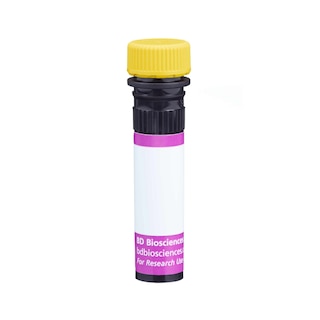
BD PharmingenTM Purified Rat Anti-Human GM-CSF
554503 ·
BDB Bioscience · Cat: 554503
Reach 3+ verified distributors
Manufacturer product page
bdbiosciences.com/en-us/products/reagents/flow-cytometry-rea...
Description
Expression of GM-CSF by stimulated human peripheral blood mononuclear cells PBMC . Ficoll-separated human PBMC were stimulated with Purified NA/LE Mouse Anti-Human CD3 1 mug/ml final concentration; Cat. No. 555329 , recombinant human IL-2 10 ng/ml final concentration; Cat. No. 554603 and recombinant human IL-4 10 ng/ml final concentration; Cat. No. 554605 for 2 days. The cells were subsequently cultured in medium containing recombinant human IL-2 and recombinant human IL-4 for 3 days. Finally, the cells were harvested and stimulated for 6 hours with PMA 50 ng/ml final concentration; Sigma, Cat. #P-8139 and calcium ionophore A23187 250 ng/ml final concentration; Sigma, Cat. #C-9275 in the presence of BD GolgiStop 2 muM final concentration; Cat. No. 554724 . The cells were harvested, stained with PE-Cy5 Mouse Anti-Human CD4 Cat. No. 555348 , fixed, permeabilized, and subsequently stained with 0.25 mug of PE Rat anti-Human GM-CSF Cat. No. 554507 antibody by using the intracellular staining protocol see Left panel . To demonstrate specificity of staining, the binding of PE Rat Anti-Human GM-CSF was blocked by preincubation of the antibody conjugate with recombinant human GM-CSF 0.1 mug; Cat. No. 550068; see Center panel and by preincubation of the fixed/permeabilized cells with Purified Rat Anti-Human GM-CSF 5 mug; Cat. No. 554503; see Right panel prior to staining. The quadrant markers for the bivariate dot plot were set based on the autofluorescence control and verified using the ligand-blocking and unlabeled antibody blocking controls. Expression of GM-CSF by stimulated human peripheral blood mononuclear cells PBMC . Ficoll-separated human PBMC were stimulated with Purified NA/LE Mouse Anti-Human CD3 1 mug/ml final concentration; Cat. No. 555329 , recombinant human IL-2 10 ng/ml final concentration; Cat. No. 554603 and recombinant human IL-4 10 ng/ml final concentration; Cat. No. 554605 for 2 days. The cells were subsequently cultured in medium containing recombinant human IL-2 and recombinant human IL-4 for 3 days. Finally, the cells were harvested and stimulated for 6 hours with PMA 50 ng/ml final concentration; Sigma, Cat. #P-8139 and calcium ionophore A23187 250 ng/ml final concentration; Sigma, Cat. #C-9275 in the presence of BD GolgiStop 2 muM final concentration; Cat. No. 554724 . The cells were harvested, stained with PE-Cy5 Mouse Anti-Human CD4 Cat. No. 555348 , fixed, permeabilized, and subsequently stained with 0.25 mug of PE Rat anti-Human GM-CSF Cat. No. 554507 antibody by using the intracellular staining protocol see Left panel . To demonstrate specificity of staining, the binding of PE Rat Anti-Human GM-CSF was blocked by preincubation of the antibody conjugate with recombinant human GM-CSF 0.1 mug; Cat. No. 550068; see Center panel and by preincubation of the fixed/permeabilized cells with Purified Rat Anti-Human GM-CSF 5 mug; Cat. No. 554503; see Right panel prior to staining. The quadrant markers for the bivariate dot plot were set based on the autofluorescence control and verified using the ligand-blocking and unlabeled antibody blocking controls. Expression of GM-CSF by stimulated human peripheral blood mononuclear cells PBMC . Ficoll-separated human PBMC were stimulated with Purified NA/LE Mouse Anti-Human CD3 1 mug/ml final concentration; Cat. No. 555329 , recombinant human IL-2 10 ng/ml final concentration; Cat. No. 554603 and recombinant human IL-4 10 ng/ml final concentration; Cat. No. 554605 for 2 days. The cells were subsequently cultured in medium containing recombinant human IL-2 and recombinant human IL-4 for 3 days. Finally, the cells were harvested and stimulated for 6 hours with PMA 50 ng/ml final concentration; Sigma, Cat. #P-8139 and calcium ionophore A23187 250 ng/ml final concentration; Sigma, Cat. #C-9275 in the presence of BD GolgiStop 2 muM final concentration; Cat. No. 554724 . The cells were harvested, stained with PE-Cy5 Mouse Anti-Human CD4 Cat. No. 555348 , fixed, permeabilized, and subsequently stained with 0.25 mug of PE Rat anti-Human GM-CSF Cat. No. 554507 antibody by using the intracellular staining protocol see Left panel . To demonstrate specificity of staining, the binding of PE Rat Anti-Human GM-CSF was blocked by preincubation of the antibody conjugate with recombinant human GM-CSF 0.1 mug; Cat. No. 550068; see Center panel and by preincubation of the fixed/permeabilized cells with Purified Rat Anti-Human GM-CSF 5 mug; Cat. No. 554503; see Right panel prior to staining. The quadrant markers for the bivariate dot plot were set based on the autofluorescence control and verified using the ligand-blocking and unlabeled antibody blocking controls. Show More Expression of GM-CSF by stimulated human peripheral blood mononuclear cells PBMC . Ficoll-separated human PBMC were stimulated with Purified NA/LE Mouse Anti-Human CD3 1 mug/ml final concentration; Cat. No. 555329 , recombinant human IL-2 10 ng/ml final concentration; Cat. No. 554603 and recombinant human IL-4 10 ng/ml final concentration; Cat. No. 554605 for 2 days. The cells were subsequently cultured in medium containing recombinant human IL-2 and recombinant human IL-4 for 3 days. Finally, the cells were harvested and stimulated for 6 hours with PMA 50 ng/ml final concentration; Sigma, Cat. #P-8139 and calcium ionophore A23187 250 ng/ml final concentration; Sigma, Cat. #C-9275 in the presence of BD GolgiStop 2 muM final concentration; Cat. No. 554724 . The cells were harvested, stained with PE-Cy5 Mouse Anti-Human CD4 Cat. No. 555348 , fixed, permeabilized, and subsequently stained with 0.25 mug of PE Rat anti-Human GM-CSF Cat. No. 554507 antibody by using the intracellular staining protocol see Left panel . To demonstrate specificity of staining, the binding of PE Rat Anti-Human GM-CSF was blocked by preincubation of the antibody conjugate with recombinant human GM-CSF 0.1 mug; Cat. No. 550068; see Center panel and by preincubation of the fixed/permeabilized cells with Purified Rat Anti-Human GM-CSF 5 mug; Cat. No. 554503; see Right panel prior to staining. The quadrant markers for the bivariate dot plot were set based on the autofluorescence control and verified using the ligand-blocking and unlabeled antibody blocking controls. Show More Expression of GM-CSF by stimulated human peripheral blood mononuclear cells PBMC . Ficoll-separated human PBMC were stimulated with Purified NA/LE Mouse Anti-Human CD3 1 mug/ml final concentration; Cat. No. 555329 , recombinant human IL-2 10 ng/ml final concentration; Cat. No. 554603 and recombinant human IL-4 10 ng/ml final concentration; Cat. No. 554605 for 2 days. The cells were subsequently cultured in medium containing recombinant human IL-2 and recombinant human IL-4 for 3 days. Finally, the cells were harvested and stimulated for 6 hours with PMA 50 ng/ml final concentration; Sigma, Cat. #P-8139 and calcium ionophore A23187 250 ng/ml final concentration; Sigma, Cat. #C-9275 in the presence of BD GolgiStop 2 muM final concentration; Cat. No. 554724 . The cells were harvested, stained with PE-Cy5 Mouse Anti-Human CD4 Cat. No. 555348 , fixed, permeabilized, and subsequently stained with 0.25 mug of PE Rat anti-Human GM-CSF Cat. No. 554507 antibody by using the intracellular staining protocol see Left panel . To demonstrate specificity of staining, the binding of PE Rat Anti-Human GM-CSF was blocked by preincubation of the antibody conjugate with recombinant human GM-CSF 0.1 mug; Cat. No. 550068; see Center panel and by preincubation of the fixed/permeabilized cells with Purified Rat Anti-Human GM-CSF 5 mug; Cat. No. 554503; see Right panel prior to staining. The quadrant markers for the bivariate dot plot were set based on the autofluorescence control and verified using the ligand-blocking and unlabeled antibody blocking controls. Expression of GM-CSF by stimulated human peripheral blood mononuclear cells PBMC . Ficoll-separated human PBMC were stimulated with Purified NA/LE Mouse Anti-Human CD3 1 mug/ml final concentration; Cat. No. 555329 , recombinant human IL-2 10 ng/ml final concentration; Cat. No. 554603 and recombinant human IL-4 10 ng/ml final concentration; Cat. No. 554605 for 2 days. The cells were subsequently cultured in medium containing recombinant human IL-2 and recombinant human IL-4 for 3 days. Finally, the cells were harvested and stimulated for 6 hours with PMA 50 ng/ml final concentration; Sigma, Cat. #P-8139 and calcium ionophore A23187 250 ng/ml final concentration; Sigma, Cat. #C-9275 in the presence of BD GolgiStop 2 muM final concentration; Cat. No. 554724 . The cells were harvested, stained with PE-Cy5 Mouse Anti-Human CD4 Cat. No. 555348 , fixed, permeabilized, and subsequently stained with 0.25 mug of PE Rat anti-Human GM-CSF Cat. No. 554507 antibody by using the intracellular staining protocol see Left panel . To demonstrate specificity of staining, the binding of PE Rat Anti-Human GM-CSF was blocked by preincubation of the antibody conjugate with recombinant human GM-CSF 0.1 mug; Cat. No. 550068; see Center panel and by preincubation of the fixed/permeabilized cells with Purified Rat Anti-Human GM-CSF 5 mug; Cat. No. 554503; see Right panel prior to staining. The quadrant markers for the bivariate dot plot were set based on the autofluorescence control and verified using the ligand-blocking and unlabeled antibody blocking controls.
More from BDB Bioscience