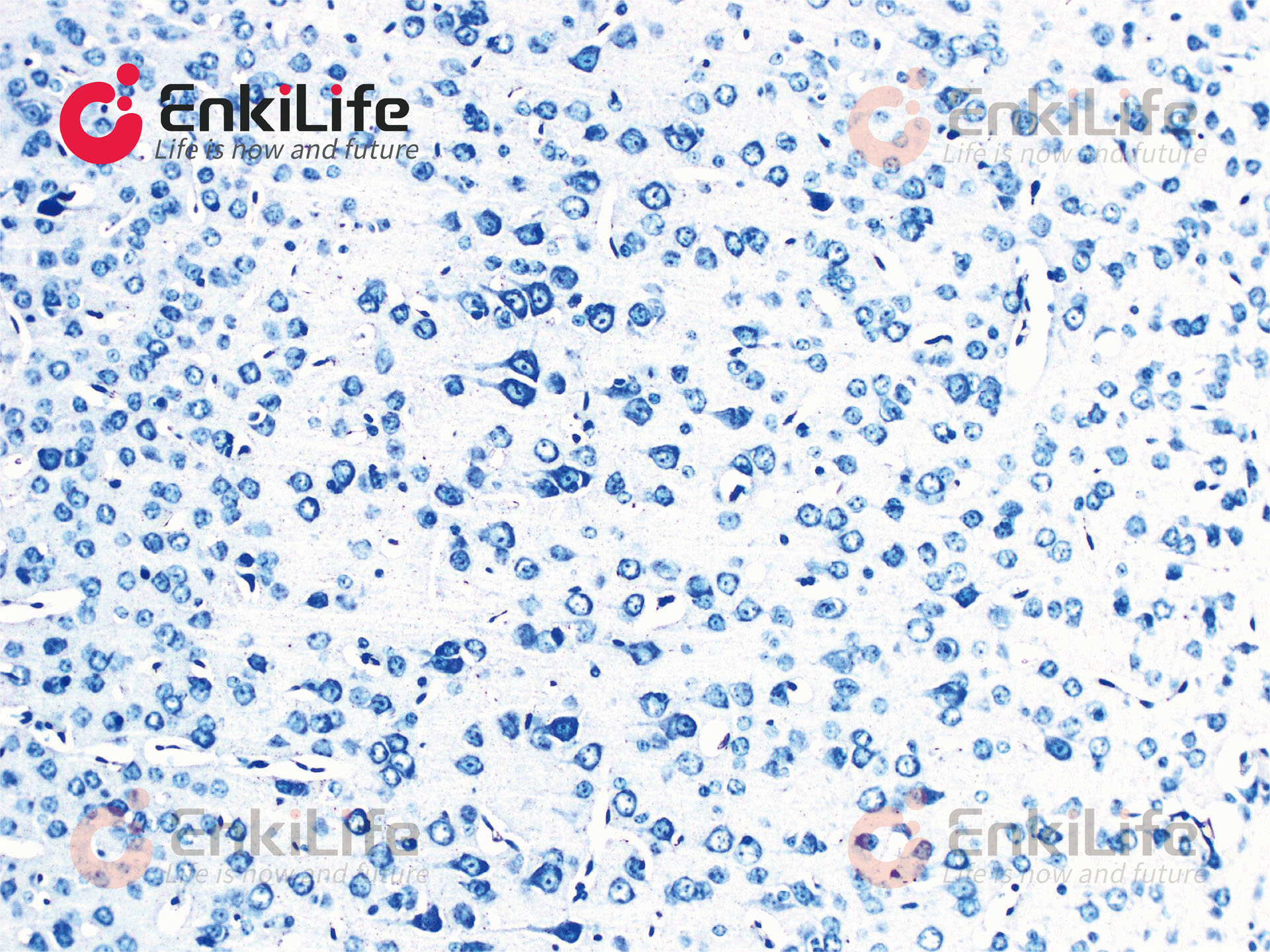
Nissl Staining Kit,Toluidine Blue Method
RA20098-100mL · 100mL
Verified Partner
Sign in to see pricing and request a quote.
Brand Store Product
No external links. Get your quote directly here.

Description
EnkiLife provide Nissl Staining Kit,Toluidine Blue Method. The neuronal cell body includes a large nucleus with a wrinkled nuclear membrane, sparse chromatin, and a prominent nucleolus. In the cytoplasm of the cell body, there are Nissl granules, which represent the rough endoplasmic reticulum and exhibit a characteristic basophilic punctate pattern in many neurons. Nissl granules can be demonstrated by various stains such as neutral red, methylene blue, toluidine blue, and methyl violet. Variations in staining, pH, and differentiation time allow some stains to highlight only Nissl substance, while others also show neuronal nuclei and neuroglia. Nissl bodies (or Nissl granules) are triangular or oval granular structures distributed in the cytoplasm of neurons. They can be stained purple-blue by basic dyes such as thionin, methylene blue, toluidine blue, and cresyl violet. All neurons contain Nissl bodies, but their shape, quantity, and distribution vary. Nissl bodies are also present in dendrites but not in axons or the axon hillock. Their appearance changes with the physiological state of the neuron. Nissl bodies are important sites for protein synthesis; when neurons are stimulated, Nissl bodies in the cell body decrease significantly.EnkiLife Nissl Staining Solution (Toluidine Blue Method) uses toluidine blue as the core dye. It is suitable for Nissl body staining in paraffin-embedded tissue sections. The presence or disappearance of Nissl bodies is an important indicator of whether neurons are damaged. In cases such as encephalitis, cerebral ischemia, or axonal reaction, Nissl bodies may dissolve or disappear.Application: Nissl Staining stain
More from EnkiLife